X-Ray
X-ray is a quick and painless imaging technique that uses
electromagnetic radiation to view the inside of the body, especially bones
What is X-RAY
X-ray is a widely used diagnostic imaging technique that employs high-energy electromagnetic waves to create images of the inside of the body. When X-rays pass through the body, different tissues absorb the rays in varying amounts—dense materials like bones absorb more and appear white on the image, while softer tissues absorb less and appear in shades of gray or black. This contrast allows doctors to examine bones, joints, and some internal organs for abnormalities.
X-rays are commonly used to detect bone fractures, infections, lung conditions, dental issues, and tumors. The procedure is usually quick, non-invasive, and painless. Although it involves exposure to a small amount of ionizing radiation, modern X-ray equipment is designed to use the lowest dose possible while still producing accurate images. Protective measures, like lead aprons, are often used to minimize exposure.

What is an MRI Used For?

Examine organs like the kidneys, liver, and digestive system

detecting fractures, dislocations, arthritis.

Help diagnose pneumonia, lung cancer, and other respiratory issues

Enlarged heart (cardiomegaly), heart failure symptoms, fluid in lungs.

Detect intestinal blockages, swallowed objects, kidney stones.

Cavities, impacted teeth, infections, bone loss, misalignment.

Our Scan Process
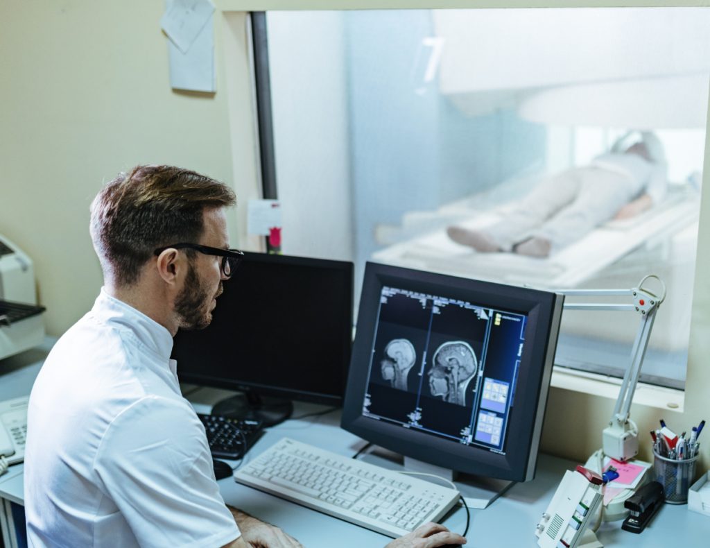
An X-ray scan is a quick, non-invasive imaging technique that uses a small dose of ionizing radiation to produce images of the internal body parts.
Preparation
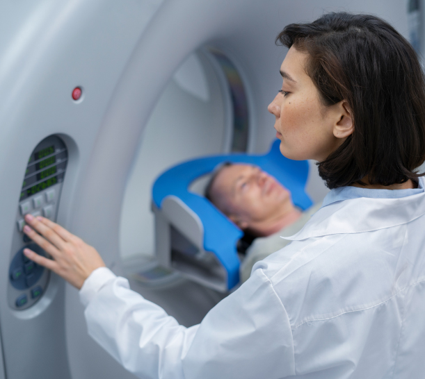
The patient is positioned depending on the area to be scanned. proper alignment ensures the correct angle and clarity of the image.
Scanning
The X-ray machine emits a controlled burst of radiation and it passes through the body
Post-Scan
An image is captured on film or digitally on a detector.
